Published June 2008
The images below illustrate Mira's MaxEnt processing applied to an inherently fuzzy image produced by Neutron Scattering Radiography. One distinction of neutron imaging is that neutrons are not affected by an electric field, so they penetrate far deeper through a conductor such as metal, while being highly attenuated by low-Z atoms and materials such as water. In contrast with X-ray images, neutron images are not sharply focused and require software processing to restore detail. The pictures below show how MaxEnt is used to improve the spatial resolution of neutron radiographs. The same technique can be applied to other imaging applications.
|
|
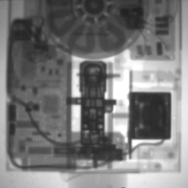
Original ImageWhat familiar object is shown in this picture? Answer: The internal details of a floppy disk drive, as imaged using Neutron Radiography. Neutrons supplied by a nuclear reactor are used like traditional X-rays to provide an internal view of this metal shrouded object. But unlike X-rays, the neutron beam is not sharply focused and there is an inherent fuzziness to the process. How can we get a sharper view? |
|
|
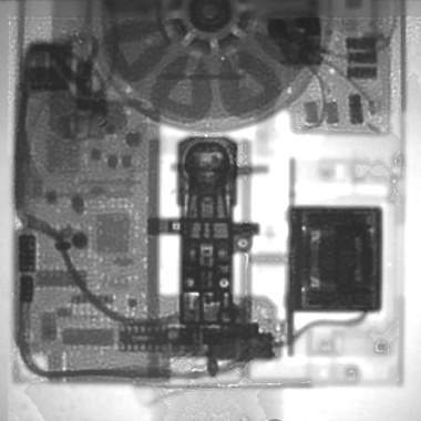
MaxEnt Processed ImageIf you've taken apart a floppy disk drive, this view probably looks familiar. In this processed image, there is an increase in overall sharpness, but there also appear new details you don't see in the original image. And they are real. Solder pads, traces, and IC sockets on the circuit board become clearly visible after Maximum entropy processing, yet are virtually invisible in the original image. |
Images courtesy Dr. Burkhard Schillinger, Technical University, Munich